
Google DeepMind เปิดตัว SynthID Bio ระบบลายน้ำสำหรับโปรตีน AI
วันนี้เรามีการเปิดตัว SynthID Bio ซึ่งเป็นเทคโนโลยีล้ำสมัยสำหรับสร้างลายน้ำให้กับโปรตีนที่ออกแบบด้วย AI โดยยังคงรักษาประสิทธิภาพการทำงานทางชีวภาพของโปรตีนเหล่านั้นไว้ได้อย่างสมบูรณ์
SynthID Bio จะทำหน้าที่ฝังลายน้ำที่มองไม่เห็นแต่สามารถตรวจสอบได้ลงในงานออกแบบทางชีวภาพโดยตรง ไม่ว่าจะเป็นลำดับโปรตีนที่สร้างโดย AI หรือโครงสร้าง 3 มิติที่ถูกพยากรณ์ไว้ก็ตาม เพื่อระบุที่มาของข้อมูลได้อย่างแม่นยำ
จากการทดสอบในห้องปฏิบัติการกับโปรตีนเป้าหมายหลากหลายชนิด พบว่างานออกแบบที่มีลายน้ำมีประสิทธิภาพและความหลากหลายตามธรรมชาติทัดเทียมกับเวอร์ชันที่ไม่มีลายน้ำ ซึ่งถือเป็นการสร้างชั้นข้อมูลต้นทาง (provenance layer) ที่สำคัญในการเสริมสร้างความปลอดภัยทางชีวภาพและรักษาความถูกต้องของฐานข้อมูลทางวิทยาศาสตร์แบบเปิด
คุณสามารถอ่านบทความบล็อกฉบับเต็มและงานวิจัยได้ที่ Google DeepMind website
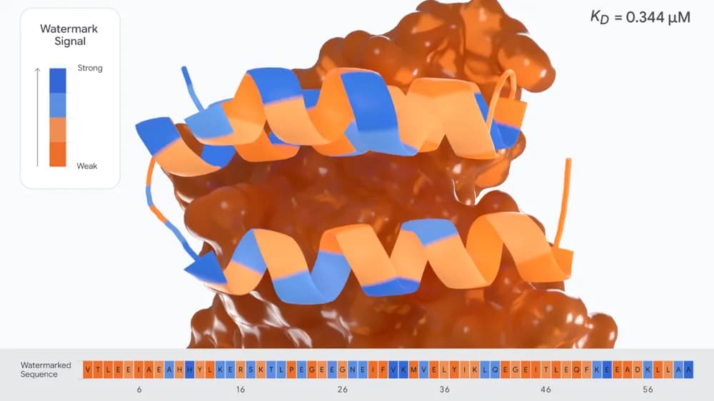
ภาพแสดงโครงสร้างพยากรณ์ของโปรตีนยึดเกาะ VEGF-A ที่มีการฝังลายน้ำ โดยสีที่ปรากฏบนกรดอะมิโนแต่ละตัวจะระบุถึงสัญญาณของลายน้ำที่ฝังอยู่
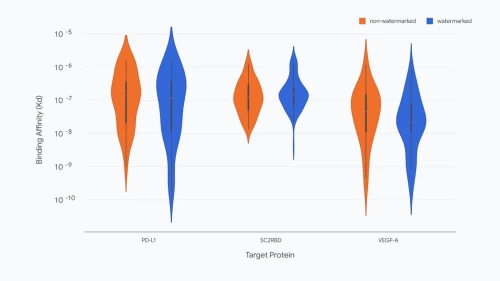
แผนภูมิเปรียบเทียบค่าความสัมพันธ์ในการยึดเกาะ (Binding affinity) หรือค่า KD ระหว่างโปรตีนที่ไม่มีลายน้ำและที่มีลายน้ำใน 3 กลุ่มเป้าหมาย ซึ่งค่าที่ต่ำกว่าจะสะท้อนถึงประสิทธิภาพการยึดเกาะที่แข็งแกร่งกว่า
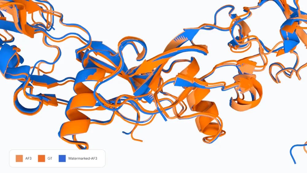
การเปรียบเทียบใน 7PPA แสดงให้เห็นถึงโครงสร้างที่พยากรณ์โดย AF3 (ซ้าย) โครงสร้างอ้างอิงตามความเป็นจริง (กลาง) และโครงสร้างที่มีการฝังลายน้ำ (ขวา) ซึ่งมีความสอดคล้องกันอย่างมาก
ความคิดเห็น (0)
เข้าสู่ระบบเพื่อร่วมแสดงความเห็น
สมัครสมาชิกมาเป็นคนแรกที่แสดงความเห็นกันเลยโบร